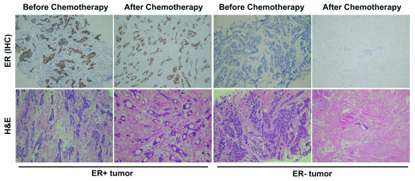
Figure 1.
Immunohistochemical staining with ER (upper panel) and H&E staining (lower panel). Stainings were performed on formalin-fixed, paraffin-embedded tissue sections of invasive ductal breast carcinoma, and photographed before and after preoperative chemotherapy treatment of EFC regimen, by using a Zeiss Axioskop40 Microscope and a Nikon E4500 camera. In ER+ tumor sample (left), tumor cells arranged in glands and clusters still showed active growth after the EFC chemotherapy treatment. Both core needle biopsy specimen and surgical specimen showed diffuse nuclear staining for estrogen receptor (diaminobenzidine chromogen). However, in ER- tumor sample (right), only a small nest of tumor cells remained survival after EFC treatment, with prominent stromal fibrosis and hyalinization. ER staining was negative in both biopsy and surgical specimens. Data are representative of at least three separate experiments. Magnification:×100.

