
Figure 3.
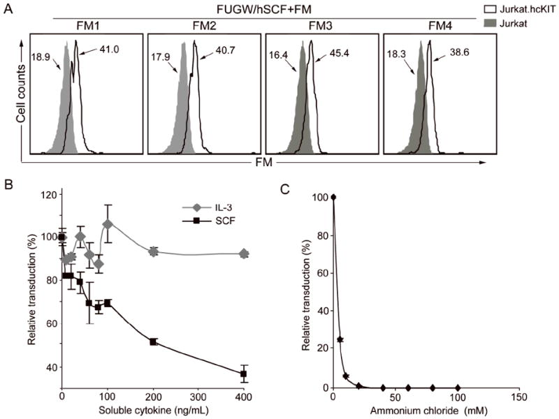
Study of the properties of in vitro targeted transduction. Fresh unconcentrated lentiviral vectors bearing hSCF and a fusogen (FUGW/hSCF + FM) were produced. (A) FACS analysis of the targeted virus cell binding by surface staining using anti-HA tag antibody against fusogen was used to detect the change in MFI due to viral vectors bound to the cells. Lentivector (FUGW/hSCF+FM) was mixed at 4°C to prevent internalization with Jurkat.hcKIT (solid line) or parental Jurkat (shaded area) as a control. (B) Effect of addition of soluble hSCF on targeted transduction. Either soluble hSCF (black square) or a control cytokine IL-3 (gray diamond) was added to the wells during transduction of Jurkat.hcKIT with FUGW/hSCF+SGM. Eight hours later, the media was replaced with fresh media and the cells placed in the incubator for four days before FACS analysis of eGFP expression. (C) Various concentrations of NH4Cl (dissolved in PBS, pH=7.4) were added into viral supernatant (FUGW/hSCF+SGM) for 8h, after which the medium was replaced with fresh medium and cells were cultured for 3 days before FACS analysis of eGFP-positive cells. The data is presented as the percentage of reduced transduction as compared to the result of transduction without treatment of either soluble cytokine (B) or NH4Cl (C).