
Figure 5.
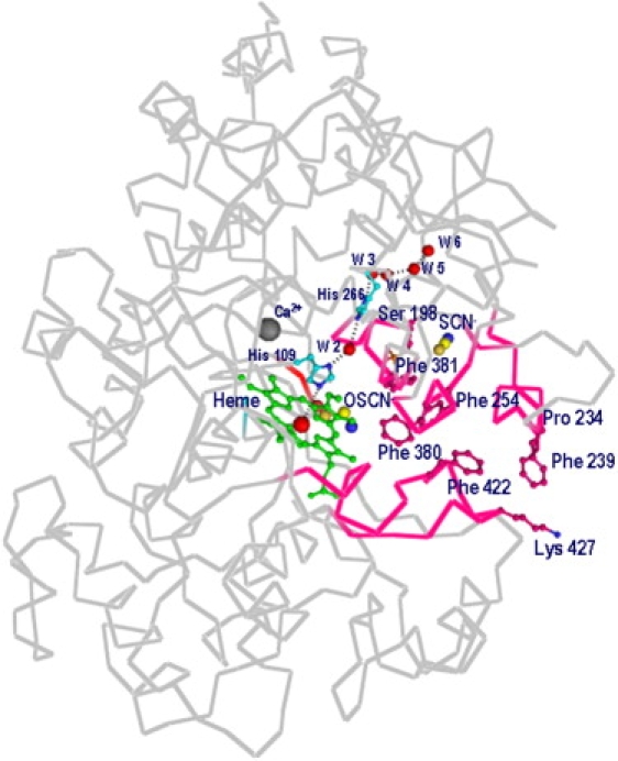
View of substrate channel connecting distal heme cavity to surface of protein. Positions of calcium and OSCN− ions, and of His-109, W2–W6, and His-266, are shown. Position of SCN− ion is also indicated. Channel walls contain predominantly hydrophobic residues, which are also indicated.