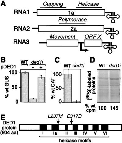
Figure 1.
Identification of DED1. (A) Schematic diagram of the BMV genome shows ORFs (open boxes), noncoding regions (single lines), tRNA-like 3′ ends (cloverleaves), and the subgenomic mRNA start site (bent arrow). (B) BMV-directed GUS expression in 1a- and 2a-expressing wt yeast, ded1i yeast, and ded1i yeast complemented with wt DED1 expressed from pDED1 is shown. (C) BMV-directed CAT expression in 1a- and 2a-expressing wt and ded1i yeast transfected with B3CAT in vitro transcripts. (D) Autoradiographs and acid-precipitable counts (below lanes) from 35S-labeled protein extracts from wt and ded1i yeast are shown. (E) Schematic diagram of Ded1p showing conserved helicase motifs (roman numerals) and substitutions in the BMV-inhibiting ded1–18 allele.

