Figure 2.
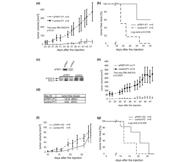
Ectopic expression of sFRP1 in MDA-MB-231 cells suppresses in vivo tumor formation. (a), (b) MDA-MB-231/secreted Frizzled-related protein 1 (sFRP1)-P1 cells and control cells (1 × 10⁶) were injected into mammary fat pads of the indicated number of Balb/c nude mice, and the tumor formation and growth were monitored: (a) average tumor volume, P < 0.01 (two-way repeated-measures analysis of variance (RM ANOVA)); (b) percentage of tumor-free mice on the indicated days after injection, P = 0.0106 (log-rank test). (c) Lysates prepared from individual tumors at the end of the experiment were monitored for Myc-tagged sFRP1 (upper panel) and for p-DVL3 and DVL3 (lower panel) by western analyses. (d) Three independent xenograft experiments using MDA-MB-231/sFRP1-P1 cells (n = 19) and control-P1 cells (n = 20) were performed and data were pooled to calculate the percentage of tumor-free mice 39 days after the injection. (e) Data from the indicated number of mice generated in two independent xenograft experiments with MDA-MB-231/sFRP1-P1 cells and control-P1 cells ((a) and Additional data file 2, Figure 2a) were pooled to yield the tumor growth curve, P < 0.0001 (two-way RM ANOVA). (f), (g) MDA-MB-231/sFRP1-P2 cells and control P2 cells (1 × 10⁶) were injected into mammary fat pads of the indicated number of Balb/c nude mice, and tumor formation and growth were monitored: (f) average tumor volume, *P < 0.01 on day 19 (Student's t test); (g) percentage of tumor-free mice on the indicated days after injection, P = 0.026 (log-rank test). (a), (d), (e) Tumor growth curves shown as the average tumor volume ± standard error.

