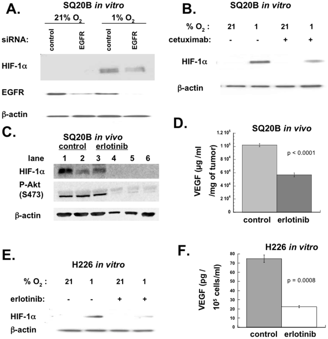
Figure 1. EGFR inhibition downregulates VEGF and HIF-1 in vitro and in vivo.
SQ20B cells were seeded at 25% confluence and cultured overnight. The next morning, cells were transfected with 150 nanomoles siRNA (either EGFR SmartPool or control non-targeted) in Optimem transfection medium using Oligofectamine. After 24 hours, the transfection medium was replaced with fresh, prewarmed culture medium. 24 hours after this (48 hours after transfection), cells were exposed to either 21% or 1% oxygen. Three hours later they were harvested and Western blotting was performed. (B) SQ20B cells were treated with 10 nM cetuximab or DMSO (control) for 16 hrs, and then exposed to either 21% or 1% oxygen. Three hours later they were harvested and Western blotting was performed. (C) Nude mice were injected subcutaneously in the flank with SQ20B cells to form xenografts. When the tumors reached a size of 100–150 mm3 (approximately 7–10 days after injection), half the mice (lanes 4–6) were started on an erlotinib-containing diet (50 mg/kg/day). After 4 days of feeding, mice were sacrificed, and the tumors were removed. Half of each tumor was lysed in protein lysis buffer, and Western blotting was performed for pAkt (S473) and HIF-1α. Each lane represents a tumor from a different mouse. (D) The other half of each tumor from (E) was homogenized in PBS, and then ELISA for VEGF was performed. VEGF level was normalized to tumor weights. Data shown represent mean values from 3 control mice and 3 erlotinib-treated mice. Error bars represent standard error of the mean. p value was obtained using Student's t-test. (E) H226 cells were treated with 10 µM erlotinib or DMSO (control) for 16 hrs, and then exposed to either 21% or 1% oxygen. Three hours later they were harvested and Western blotting was performed. (F) H226 cells were seeded then later that day they were treated with erlotinib (10 µM) or DMSO (control). 24 hours later, the media was changed and replaced with media containing 1% serum with or without erlotinib. Cells were exposed to 1% oxygen. 16 hours later, aliquots of supernatant were removed from dishes, and ELISA for VEGF was performed. ELISA values were normalized to the number of cells present. Data shown represent mean values. Error bars represent standard error of the mean. p value was obtained using Student's t-test.

