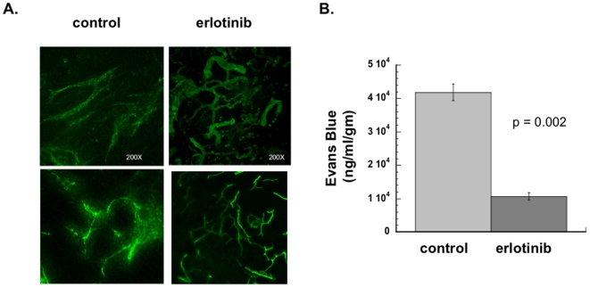
Figure 2. Erlotinib alters vascular morphology and increases vascular permeability in vivo.
(A) Nude mice were injected subcutaneously in the flank with SQ20B cells to form xenografts. When tumors reached a size of ∼1 cm in diameter, some of the mice were started on an erlotinib-containing diet (50 mg/kg/day). After 4 days of erlotinib treatment (or control diet), FITC-conjugated tomato lectin was injected via tail vein. The mice were euthanized, and then a 100 µm thick section of each tumor was viewed under confocal microscope. The projected photomicrograph (200× total magnification) of sections of tumors from 2 different erlotinib-treated and 2 control mice are shown (B) Tumors were grown as described in (A). Evans blue dye was injected intravenously. Six hours later mice were sacrificed and tumors were excised. Dye was extracted, then quantified by reading at 620 nm in a spectrophotometer. Data shown represent mean of 3 control mice and 3 erlotinib-treated mice. Error bars represent standard error of the mean. p value was obtained using Student's t-test.

