
Figure 1.
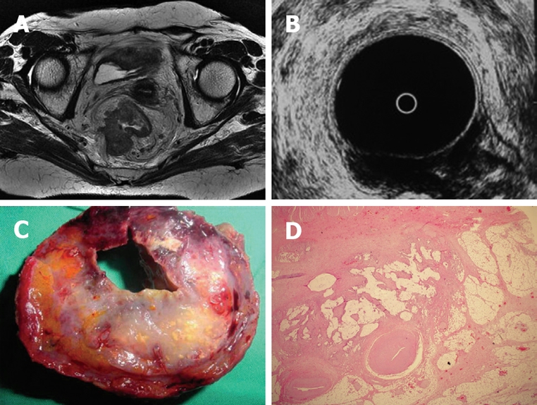
A: MRI demonstrates a large tumor passing through the muscularis propria and invading the mesorectal fatty tissue within very close proximity to the mesorectal fascia; lymph nodes are also present; The MRI prediction was a T4 tumor; B: This was predicted as a T3 tumor by ERUS examination; C: Macroscopic specimen shows that the tumor has already filled all the mesorectal fatty tissue but the mesorectal fascia is still intact; D: Pathological examination reveals that this is a T3 stage tumor.