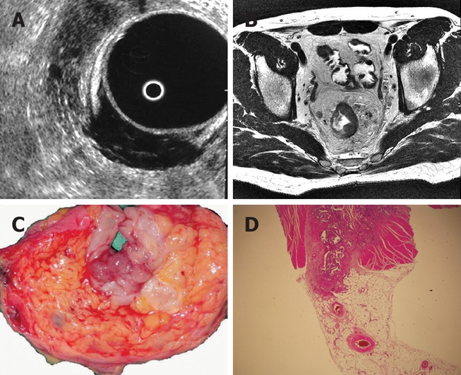
Figure 3.

A: ERUS examination shows the tumor extend to the mesorectal fat by passing beyond the muscularis propria. A lymph node is also seen; B: MRI defines the tumor as violating the muscularis propria and extending to the mesorectal fatty tissue. Lymph nodes are seen; C: Operation specimen confirms mesorectal invasion; D: Pathology specimen demonstrates tumor cells invading the mesorectum which is indicative of a T3 tumor.
