
Figure 1.
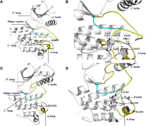
(A) The crystal structure of the wild-type RET kinase in the active form (PDB entry 2IVS). (B) A closeup of structural environment near M918T mutation in the RET kinase. (C) The crystal structure of the wild-type MET kinase in the inactive, autoinhibited form (PDB entry 2G15). (D) A closeup of structural environment near M1250T mutation in the MET kinase.