
Figure 7.
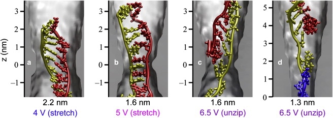
Possible conformations of hpDNA during translocation. (a) Conformation of the bases as the double helix passes by stretching/distortion through the 2.2-nm pore. (b) Conformation of the bases as the double helix passes by stretching/distortion through the 1.6-nm pore. Note the difference between the conformation here and that in panel a. (c) Conformation of the bases as the double helix unzips in the 1.6-nm pore. Note the difference in the orientation of the bases as compared to the stretching/distortion pathway. (d) Conformation of the bases as the double helix unzips in the 1.3-nm pore.