
Figure 1.
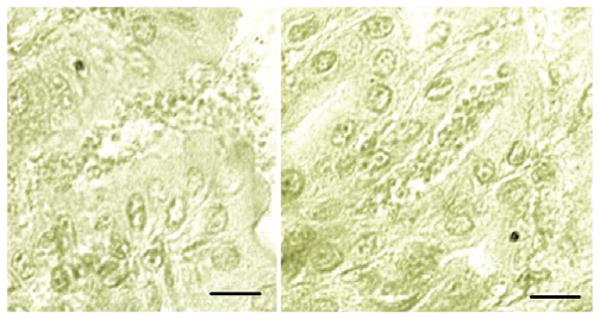
Micrographs of Tyzzer's histological sections of the stomach of an experimentally infected mouse. The information pertaining to this slide is copied from a handwritten catalogue card (# 1897), dated Jan 16, 1908, and entitled ‘Cryptosporidium feeding experiment’. The above micrographs were taken by Xiaochuan Feng, Tufts Cummings School of Veterinary Medicine, from an archive slide containing the gastric section of a mouse experimentally fed infected stomach contents and gastric mucus and sacrificed 14 days later. Note the small forms outpouring from an open pit onto the gastric surface (left) and a pit filled with parasite forms (right). There was no reference to fixative or stain used. Scale bar = 20 μm.