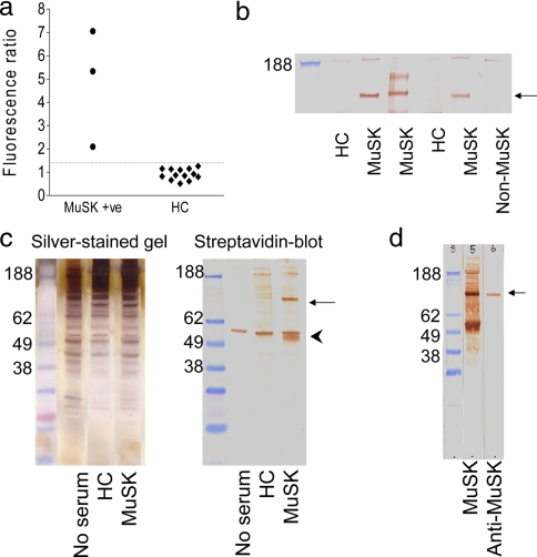
Fig. 2.
Evidence for a specific membrane antigen and its immunoprecipitation by MuSK-antibody positive IgG. a, three MuSK-MG plasmas displayed binding to TE671 cells (median fluorescence intensity) greater than the mean + 2 S.D. of the binding of control sera (dotted line). b, incubation of a surface-biotinylated TE671 cell extract with any one of three MuSK-MG plasmas resulted in the immunoprecipitation of a 90-kDa band of biotinylated protein. This band was not present when immunoprecipitation was performed using two healthy control subject sera or using a MuSK-seronegative MG patient plasma. c, the immunoprecipitation technique was refined to reduce nonspecific binding by incubation of intact TE671 cells with plasma followed by washing before cell extraction. The silver-stained gel contained a large number of protein bands matched between the patient, healthy control, and blank immunoprecipitates. On the blot, which was probed with HRP-conjugated streptavidin, there was a strong band at 90 kDa (arrow), which was exclusive to the MuSK-MG immunoprecipitate, and a band around 55 kDa present in all lanes (arrowhead) that must have been nonspecifically captured. d, a goat anti-MuSK antibody bound to a 90-kDa protein band derived from unbiotinylated TE671 cells, which had been immunoprecipitated using a MuSK-MG plasma. This band was of the same molecular mass as the band derived from surface-biotinylated TE671 cells, which had been immunoprecipitated using the same MuSK-MG plasma. HC, healthy control; +ve, positive.

