Fig. 6.
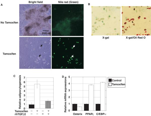
Tamoxifen-induced ALK5 inactivation increased adipocytogenesis in primary calvarial cell cultures. (A) Representative light and fluorescent microscopic images of Nile Red (green signal) staining of primary calvarial cell cultures in the absence or presence of tamoxifen at day 18 after osteoinduction. Droplets observed in light microscopy (left, black arrows) were positive for Nile Red lipophilic fluorescent dye (right, white arrows). (B) Microscopic pictures of X-gal/Oil Red O-staining of primary calvarial cell cultures in the absence or presence of tamoxifen at day 20 after osteoinduction. Note that many Oil Red O-positive cells were also positive for X-gal staining. (C) Adipocytogenesis was quantified by the Nile Red positive area as described in Materials and Methods. Data represent means ± SD. Inactivation of ALK5 resulted in enhanced adipocytogenesis of calvarial cells. (D) Real-time quantitative RT-PCR for the osteoblast-specific osterix and adipocyte-specific differentiation markers PPARγ and C/EBPα. cDNA, prepared using mRNA from calvarial cells on the 6th day of the osteogenic culture, was subjected to real-time quantitative PCR. Osteogenic marker expression was downregulated while adipocyte marker expression was upregulated in the presence of tamoxifen.

