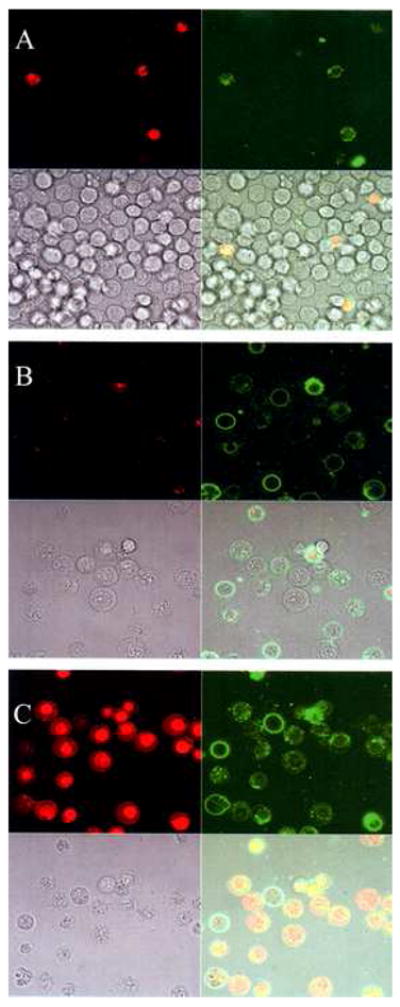
Fig 1.

Phase and fluorescence (Annexin (+) and EthD-1 (−) ) microscope images of electric field induced PS externalization in B-cells. (A) Control – No Electric field. (B) Electroporated B-cells. E ~ 2.1 kV/cm, ~ 200 μSec pulse. (C) Addition of 0.2% Triton X-100 to the sample shown in (B) to permeabilize the membrane.
