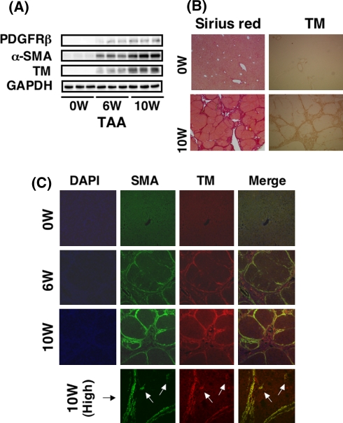
Fig. 2.
Expression of tropomyosin in fibrotic livers. The expression of tropomyosin in the fibrotic liver was determined by immunoblot and immunohistochemistry. A Whole-liver homogenates were subjected to SDS-PAGE, transferred onto the membrane, and successively immunoreacted with PDGFR-β, α-SMA, or tropomyosin. Note that tropomyosin is induced in the liver of rats treated with TAA time dependently after starting injection in a similar manner to the expression of PDGFR-β and α-SMA. B Histology. Prominent liver fibrosis is observed in the liver of rats treated with TAA for 10 weeks by Sirius red staining. Tropomyosin expression is clear along the septa. C Fluorescent immunohistochemistry of tropomyosin and α-SMA. Double immunostaining was performed in the liver of rats treated with TAA for 6 and 10 weeks. Note that both proteins always colocalize and are expressed strongly at the site between noninjurious parenchyma and septa (magnification ×100). Activated HSCs (arrows) that were present close to the septum and positive for α-SMA also expressed tropomyosin (magnification ×400)

