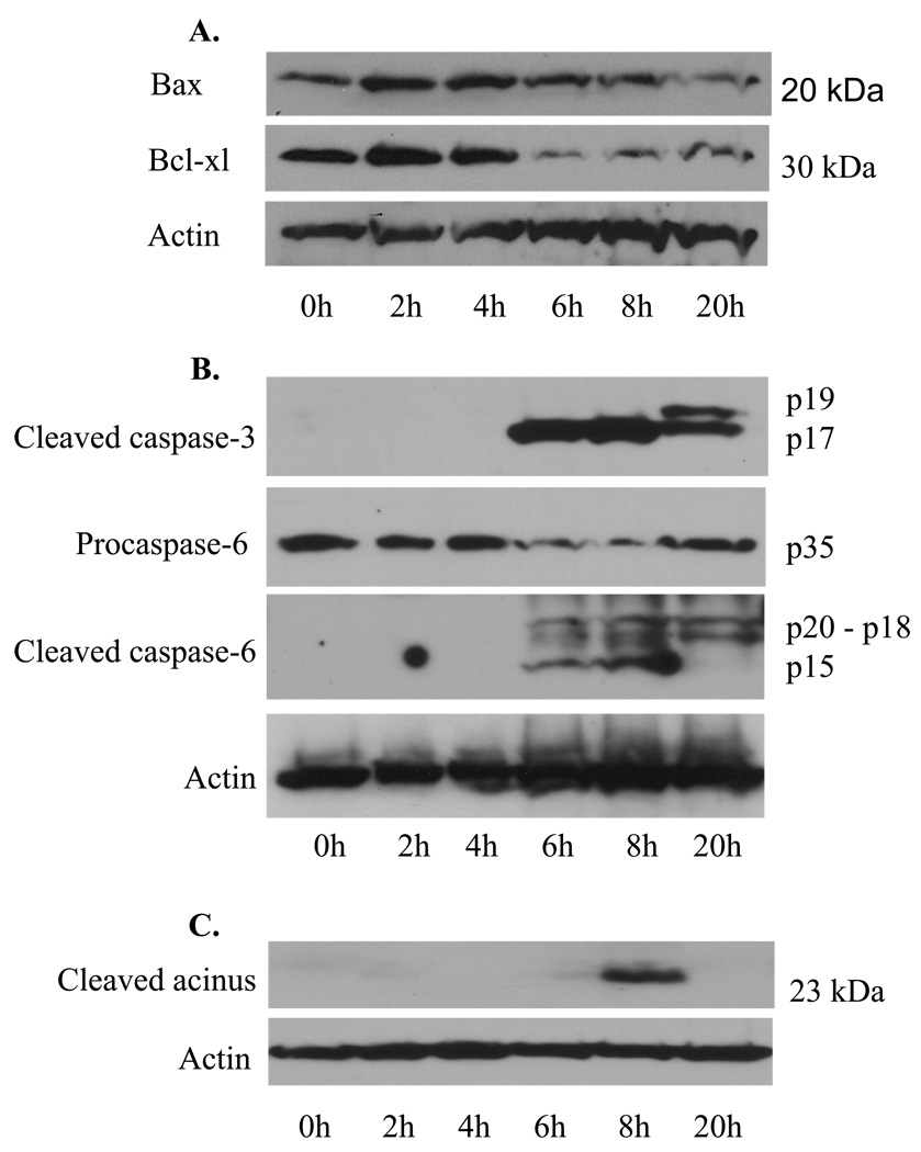
Figure 3. Expression of apoptotic mediators during the development of experimental PF.
At various time points post PF1 IgG injection, mouse skin specimens were harvested, pooled (n=2, each time point), and extracted. The extracts were subjected to Western blot analysis using a polyclonal antibody for Bax, a monoclonal antibody to Bcl-xl (A), a monoclonal antibody specific for the cleaved caspase-3, a polyclonal antibody for cspase-6 (cleaved and procaspase-6) (B), or a polyclonal antibody against the cleaved acinus (23 kDa) (C). β-actin was detected for protein loading controls. As shown, the level of pro-apoptotic factor Bax was slightly increased at 2 and 4 h, whereas the anti-apoptotic factor Bcl-xl expression was reduced markedly at 6, 8, and 20 h. The cleaved (activated) of caspase-3 and -6 were detected by 6, 8, and 20 h after IgG injection. Acinus cleavage was revealed transiently at 8 h.

