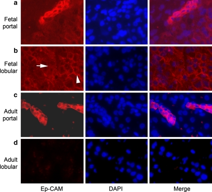
Fig. 4.
Immunofluorescence localization of hepatic Ep-CAM. Sequential images of tissues stained for Ep-CAM (red color) and DAPI (blue) with merged images in panels on extreme right are shown. Panels in a and b show 16-week-old fetal liver and panels in c and d show adult liver. Note expression of Ep-CAM at high levels throughout cell membranes in ductal plate cells in fetal liver (a) and bile duct cells in adult liver (b). In hepatoblasts, Ep-CAM expression was less intense, and Ep-CAM was distributed in a focal manner (arrow, b) in cell membrane (arrowhead, b). Adult hepatocytes did not express Ep-CAM. Original magnification, 400×

