Figure 5.
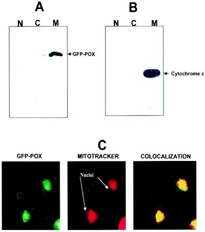
Recombinant GFP-proline oxidase localizes to mitochondria. Cells were subfractionated into nuclei (N), cytoplasmic/membrane (C), and mitochondrial (M) fractions. (A and B) A portion of each subcellular fraction (5 μg protein) was solubilized in SDS gel loading buffer and immunoblotted with a polyclonal GFP antibody (A) or a monoclonal cytochrome c antibody (B). (C) Confocal microscopy was used to visualize the localization of GFP-proline oxidase in transfected H1299 cells (GFP-POX; green fluorescence). These same cells were counterstained with MitoTracker Red (red fluorescence) to visualize the localized concentration of mitochondria in the cytoplasm. The right panel shows the simultaneous excitation of GFP-proline oxidase and MitoTracker Red in the same field as that shown in the left and middle panels, which yielded a yellowish-red fluorescence at areas of colocalization of GFP-POX and MitoTracker Red. Filtered control analyses indicated that there was no contamination of the red fluorescence window by the green fluorescence signal or of the green fluorescence window by the red fluorescence signal.

