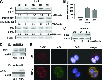
Figure 4.
p-p38 is a direct interacting partner of DAPK during TNFα-induced apoptosis. A: Lysates of HCT116 cells subjected to TNFα were analyzed by Western blotting using anti-p38, anti-p-p38, anti-ERK1/ERK2, anti-p-ERK1/ERK2, anti-JNK, anti-p-JNK, or anti-β-actin. Untreated HCT116 cells (ctrl) served as a control. B: HCT116 cells (ctrl or subjected to TNFα) were assayed for p38 MAPK activity; the control was adjusted to one. C: Lysates of HCT116 cells treated with supernatants from human actM isolated from PBMCs were analyzed by anti p-p38 and anti-β-actin Western blotting. Untreated HCT116 served as a control. D: HCT116 cells subjected to TNFα were lysed, and DAPK or p-p38 proteins were immunoprecipitated (IP) using anti-DAPK or anti-p-p38. Precipitates were analyzed by Western blotting for the presence of p-p38 and DAPK. E: TNFα-induced co-localization of DAPK and p-p38 after 6 hours was determined by fluorescence immunolabeling analysis using anti-DAPK, anti-p-p38, and DAPI.

