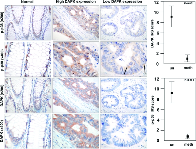
Figure 8.
DAPK co-localizes with p-p38 in human colorectal carcinoma. Immunohistochemical analysis of DAPK and p-p38 in normal colonic mucosa and colorectal carcinoma tissue with and without DAPK expression was detected by anti-DAPK and anti-p-p38. un, umethylated tumors; meth, methylated tumor (Microscope: Zeiss Axioscope 50; camera: Nikon coolpix 990; magnification: ×200, ×400). Arrows show single tumor cells expressing p-p38. Statistical analysis of differences in immunohistochemical DAPK and p-p38 expression (Student’s t-test).

