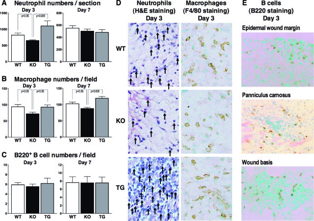
Figure 2.
Inflammatory cell recruitment in wounded skin from wild-type (WT), CD19−/− (KO), and CD19Tg (TG) mice at 3 and 7 days after injury. A: Numbers of neutrophils per section were determined by counting H&E-stained sections under the microscope. Numbers of F4/80+ macrophages (B) and B220+ B cells (C) per field (0.07 mm2) were also counted under the microscope. D: Representative histological skin sections from wild-type, CD19−/−, and CD19Tg mice at 3 days after wounding (original magnification, ×400). Arrows represent neutrophils. E: Representative histological skin sections from wild-type mice at 3 days after wounding (magnification = original ×400). Each histogram shows the mean ± SEM values obtained from 10 mice (10 wounds) in each group.

