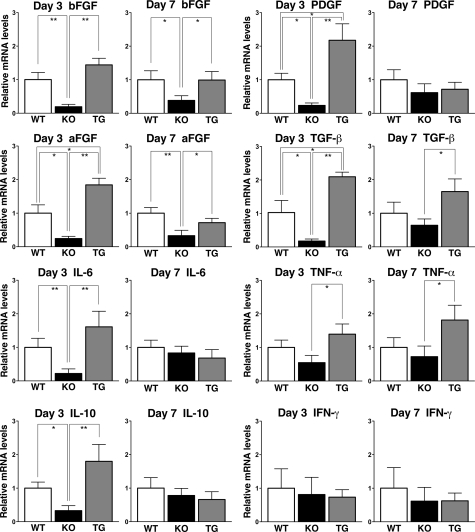
Figure 3.
Cytokine production in the wounded skin tissue of wild-type (WT), CD19−/− (KO), and CD19Tg (TG) mice. Relative mRNA expression of bFGF, aFGF, IL-6, IL-10, PDGF, TGF-β, TNF-α, and IFN-γ was quantified by real-time PCR and normalized relative to endogenous glyceraldehyde-3-phosphate dehydrogenase levels. Each histogram shows the mean ± SEM values obtained from 10 mice (10 wounds) in each group. *P < 0.05, **P < 0.01.

