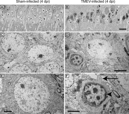
Figure 10.
EM evidence that hippocampal pyramidal neurons undergo apoptosis during acute TMEV infection. Electron microscopy was used to examine cellular morphology in the CA1 layer of the hippocampus. Hippocampal thick sections were collected at 4 dpi from sham-infected (A,C,E) and TMEV-infected (B,D,F) mice and stained with Toluidine Blue O to identify the CA1 region. Thin sections of CA1 were then prepared for EM analysis. Note the considerable difference in pyramidal cell layer architecture and the apoptotic morphology of pyramidal neurons between sham- (A) and TMEV-infected (B) animals at low magnification. Higher magnification showed the morphology of normal pyramidal neurons in sham-infected mice (C,E). In sections from TMEV-infected mice (D,F) the majority of CA1 neurons showed a shrunken morphology, cellular blebbing (arrow in F), and chromatin condensation along the nuclear membrane (arrowhead in F), consistent with apoptosis. In addition, vacuolization of the area surrounding the dying neuron (D,F) was consistent with the vacuolization seen by histological analysis and light microscopy. Scale bars: 20 μm (in B and applies to A); 5 μm (D and applies to C); 2.5 μm (E); 2.5 μm (F).

