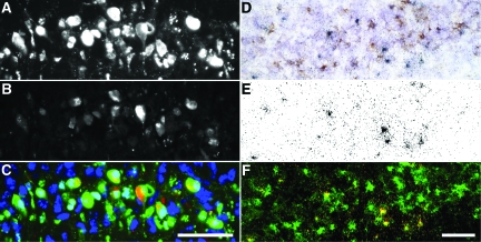
Figure 12.
Death of CA1 pyramidal neurons is independent of direct infection with the TMEV virus. Colocalization of TUNEL immunostaining (A; green in C) and TMEV antigen immunostaining (B; red in C) revealed the presence of many dying neurons that were not infected with the virus (green-only cells in C) at 4 dpi. This observation was confirmed by the extensive absence of colocalization between TUNEL immunohistochemistry (D; pseudocolored green in F) and in situ hybridization for viral RNA (grains in E; pseudocolored red in F) at 4 dpi. While several cells are both infected and dying (yellow in F), the majority of dying neurons are not infected (green-only in F). DAPI is shown in blue in (C). Scale bar: 50 μm (C and refers to A–C); 50 μm in (F and refers to D–F).

