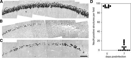
Figure 7.
The majority of CA1 pyramidal neurons are lost by 7 dpi. Hippocampal sections at −1.7 mm from Bregma were collected from uninfected mice (A) and mice at 7 dpi (B and C) and stained for the neuron-specific marker NeuN. Montages of images collected at ×60 reveal that the high density of CA1 neurons present in uninfected controls (A) is attenuated following infection (B and C). D: Individual fields were subjected to an automated thresholding and counting routine to determine the number of NeuN-positive cells in CA1. While greater than 90 NeuN-positive cells were found per field in uninfected mice (n = 20 fields from 5 mice), fewer than 20 NeuN-positive cells were observed per field in mice at 7 dpi (n = 20 fields from 5 mice), and many animals had CA1 fields devoid of NeuN-positive structures. Scale bar in (C) = 100 μm; refers to A–C.

