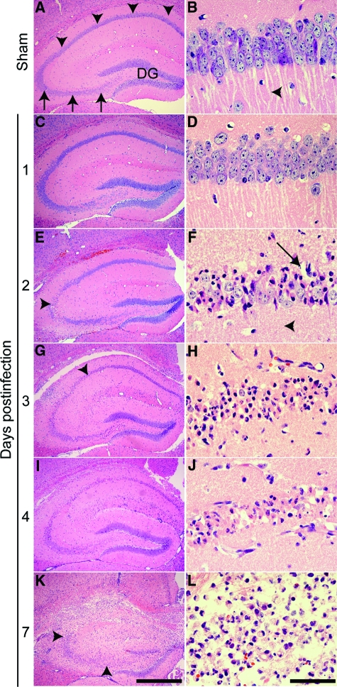
Figure 8.
TMEV infection triggers a time-dependent injury to the pyramidal layer (stratum pyramidale) of the CA1 region of the hippocampus. A, C, E, G, I, K: Low magnification H&E image of the hippocampus. B, D, F, H, J, L: Higher magnification image collected within a portion of CA1 chosen to highlight representative pathology. A and B: Sham-infected animals exhibit normal hippocampal morphology 7 days after infection. The CA1 pyramidal layer is indicated by arrowheads in (A). The CA3 pyramidal layer is indicated by arrows in (A). DG: dentate gyrus. Normal CA1 pyramidal neuron apical dendrites are marked by an arrowhead in (B). C and D: No damage was evident at 1 dpi. E and F: By 2 dpi, pyramidal neuron damage near the CA1 and CA3 junction was detectable (arrowhead in E). Higher magnification of the damaged area revealed condensed neurons, vacuolization in the pyramidal layer architecture (arrow in F), and complete loss of the large apical dendrites (arrowhead in F). G and H: This process continued at 3 dpi, with the injury spreading medially along CA1 (arrowhead in G). I and J: By 4 dpi the majority of neurons in CA1 were injured or absent. K and L: By 7 dpi there was frank destruction of the hippocampal architecture surrounding CA1 but remarkable preservation of CA3 (delineated by arrowheads in K) and the dentate gyrus. Scale bar in (K) = 500 μm; refers to A, C, E, G, I, and K. Scale bar in (L) = 50 μm; refers to B, D, F, H, J, and L. These results are representative of at least 10 mice in each of three separate experiments.

