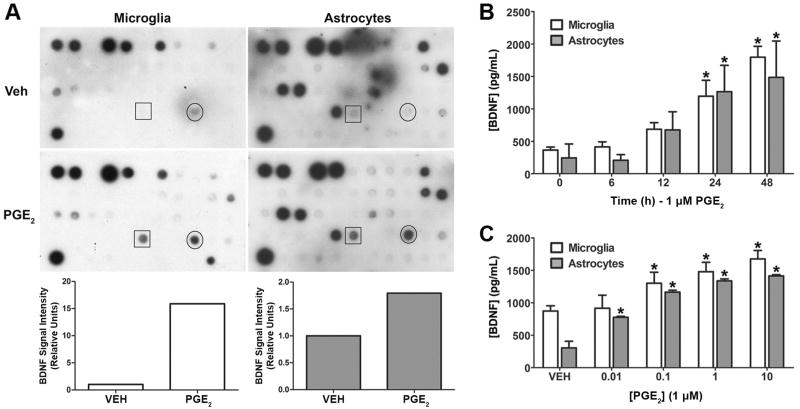
Figure 1. PGE2 stimulates the secretion of BDNF from cultured human microglia and astrocytes.
(A). PGE2 stimulated BDNF release from human microglial cells and astrocytes was initially identified by using an antibody microarray to screen a panel of human cytokines and growth factors. The arrays were incubated in conditioned media supernatants from microglia (left) and astrocytes (right) treated with vehicle or 1 μM PGE2 for 24 h. Signals from the BDNF antibody are shown in the boxes. Signals for VEGF are circled. Bar graphs show the results of densitometry analysis of the scanned autoradiograms for BDNF. BDNF signal values were normalized to the corresponding positive control values derived from the upper left spots on each array. (B). Regulation of BDNF secretion was confirmed by ELISA analysis of conditioned media from PGE2 treated microglial and astrocyte cultures. The graph shows the accumulation of BDNF in supernatants from microglial cells (open bars) and astrocytes (filled bars) treated with PGE2 for the indicated times. (C). ELISA analysis of the effect of PGE2 concentration on the secretion of BDNF from microglia and astrocytes. ELISA data are shown as the mean ± SD of triplicate measurements from a representative experiment that was repeated three times. *p < 0.05 compared to 0 h time point or vehicle control.

