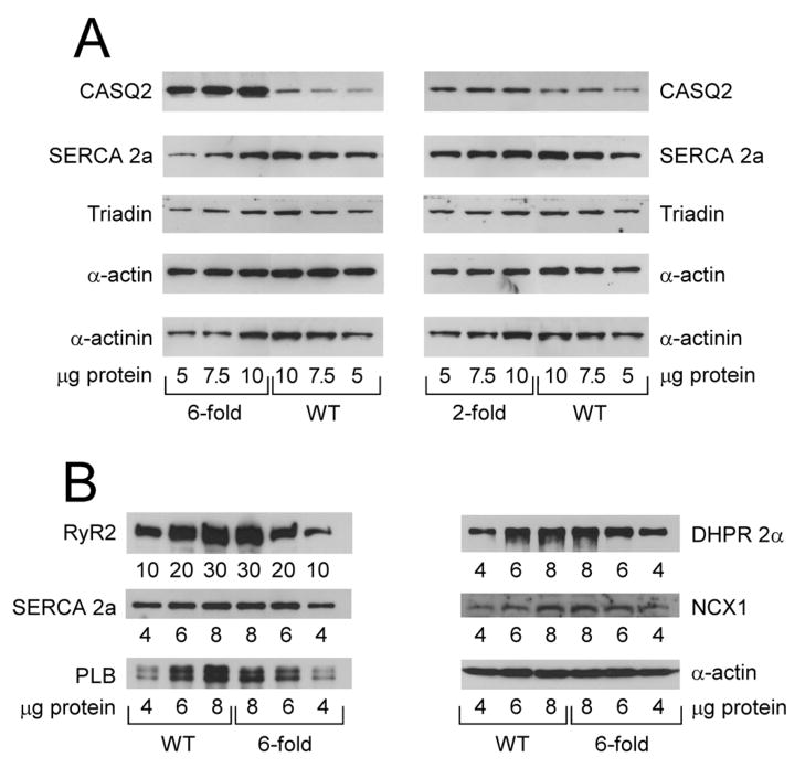
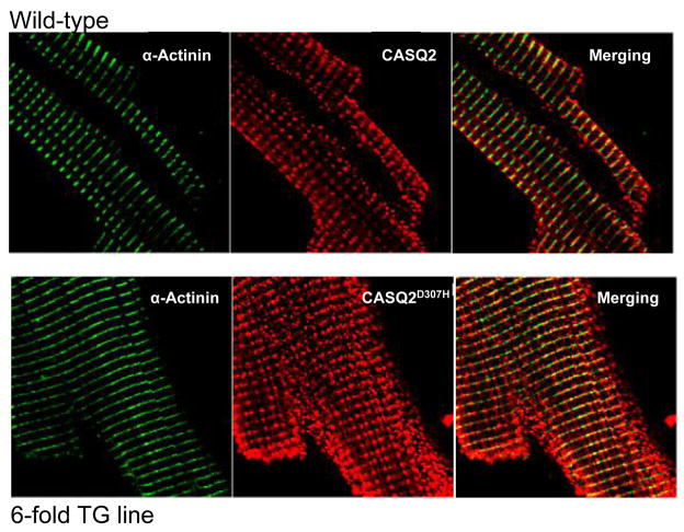
Fig. 1. (A–B) Expression of SR proteins in TG (2- and 6-fold CASQ2D307H) and WT hearts.
(A) 5–10 μg of whole heart homogenates (2- and 6-fold CASQ2D307H) were separated on 10% SDS-PAGE gels and probed with antibodies against CASQ2, SERCA 2a, triadin, α-actin and α-actinin. (B) 4–8 μg (10–30 μg for RyR2) of whole heart homogenates (6-fold CASQ2D307H) were separated on SDS-PAGE gels and probed with antibodies against RyR2 (4–20% gradient gel), SERCA 2a (10% gel), phospholamban (PLB; 15% gel), L-type calcium channel (DHPR 2α; 10% gel) and Na+/Ca2+ exchanger (NCX1, 10% gel). The 120-kDa full length NCX1 protein is shown 44. (C) CASQ2 immunofluorescence in TG and WT myocytes. Mice ventricular cardiomyocytes from WT littermates and TG (6-fold CASQ2D307H) were immunostained with mouse anti α-actinin (1:500) or rabbit anti-calsequestrin (1:200), followed by Alexa 488-conjugated goat anti-mouse (1:300) and Cy™3-conjugated Donkey anti-rabbit (1:300 dilution) antibodies.


