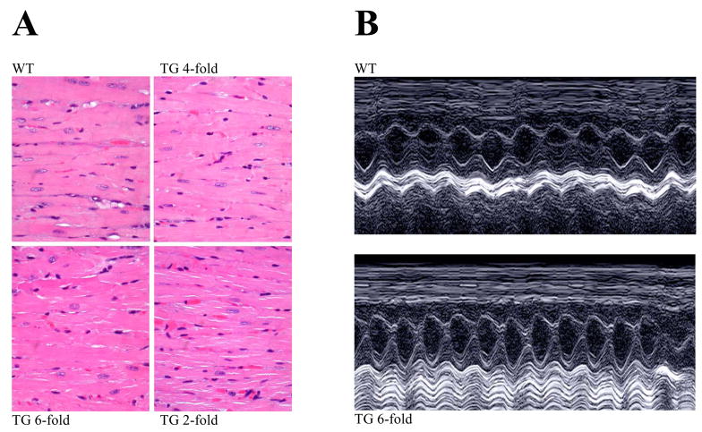
Fig. 3. TG hearts show normal morphology and function is unaltered.
(A) Hearts from TG mice (~1 year old) were subjected to standard histological analysis by staining with hematoxylin/eosin. (B) Examples of M-mode echocardiography in 4–6 month old WT and TG mice (6-fold CASQ2D307H) showing normal cardiac chamber and contractility with no evidence of structural abnormalities. Summary of data are shown in Table 1.

