Figure 1.

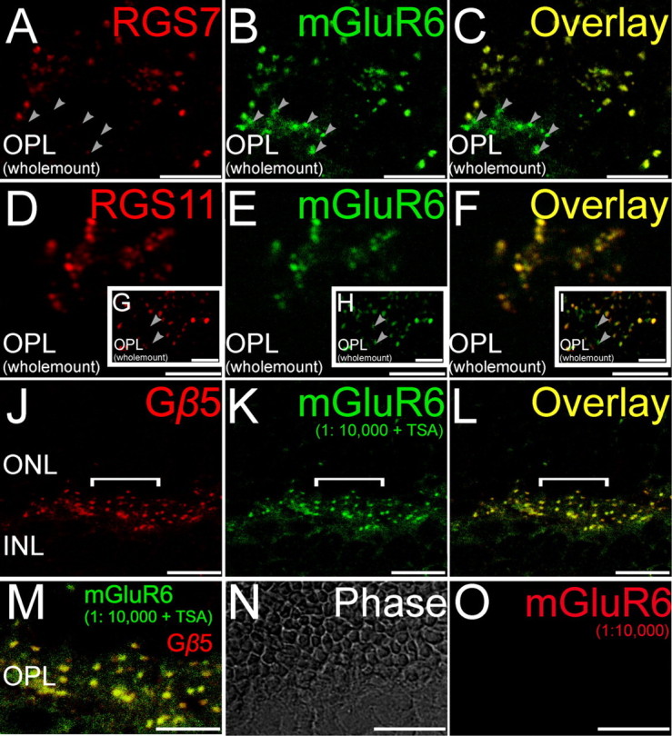
Double staining for GAP proteins and mGluR6. Double labeling using antibodies directed against RGS7, RGS11, and Gβ5 (red) and the ON bipolar cell marker mGluR6 (green). A–C, (Wholemount), Labeling for RGS7 colocalized with labeling for mGluR6. Some closely clustered dendritic tips likely representing ON cone bipolar cells were immunopositive for mGluR6 but not RGS7 (arrowheads). D–F (Wholemount), Labeling for RGS11 in the mouse retina colocalized extensively with labeling for mGluR6 especially at tightly clustered ON bipolar cell dendritic tips representing putative ON cone bipolar cells. G–I (Wholemount insets), Some isolated dendritic tips, likely representing rod bipolar cells, were immunopositive for mGluR6 but not RGS11 (arrowheads). J–L (Radial section), Labeling for Gβ5 in the mouse retina colocalized extensively with labeling for mGluR6. M (Radial section), The overlay of the region indicated by the white horizontal line in J–L is shown here in an expanded scale to show the colocalization of Gβ5 and mGluR6. N, O (Radial section), Tyramide signal amplification did not produce any visible immunolabeling at the dilution of 1:10,000 for the red channel, eliminating the possibility of cross reaction of the secondary antibodies detected in the red channel with the primary detected in the green channel. Scale bars: A–I, M–O, 5 μm; J–L, 10 μm. ONL, Outer nuclear layer; INL, inner nuclear layer.
