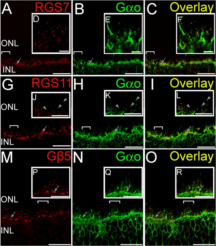
Figure 2.

Double staining for GAP proteins and Gαo. Double labeling using antibodies directed against RGS7, RGS11, and Gβ5 (red) and Gαo (green). A–C (Radial section), Whereas Gαo staining was widely distributed in ON bipolar cells, the punctuate staining for RGS7 (A) in the OPL colocalized with labeling for Gαo (B) at the dendritic tips of ON bipolar cells as seen from the overlay (C). Clusters of RGS7 staining (arrows) indicate their presence in cone bipolar cells. D–F (Insets of bracketed regions in A–C magnified), The punctuate staining for RGS11 (D) in the OPL colocalized with labeling for Gαo (E) at the dendritic tips of ON bipolar cells as seen from the overlay (F). G–I (Radial section), Clusters of RGS11 staining (arrows) indicate their presence in cone bipolar cells. J–L (Insets of bracketed regions in G–I magnified), Some rod-bipolar cell dendritic tips (arrowheads) were devoid of detectable staining for RGS11. M–O (Radial section), The punctuate staining for Gβ5 (M) in the OPL colocalized with labeling for Gαo (N) at the dendritic tips of ON bipolar cells as seen from the overlay (O). P–R (Insets of bracketed regions in M–O magnified), Clusters of Gβ5 staining (arrows) indicate their presence in cone bipolar cells. Scale bars: A–C, G–I, and M–O, 20 μm; D–F, J–L, and P–R, 5 μm. ONL, Outer nuclear layer; INL, inner nuclear layer.
