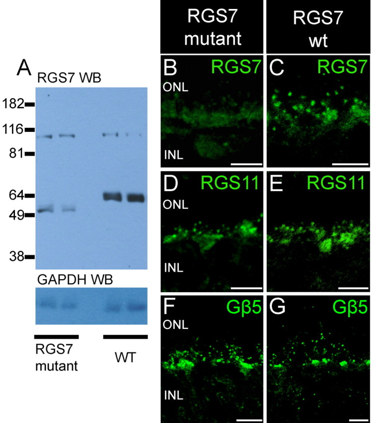
Figure 5.

Absence of full-length protein in RGS7 mutant mice. A, Top panel, Western blot analysis of retinal lysates (1/30 of a retina) from two RGS7 mutant mice and two wild-type mice using affinity purified anti-bovine RGS7 antibody R4163. The anti-RGS7 antibody detected a band at molecular weight of ∼ 58 kDa in the lanes containing retinal lysates from wild-type mice. In the lanes containing retinal lysates from RGS7 mutant mice, the anti-RGS7 antibody detected a band at a position lower than that of the wild-type RGS7 band. The signal intensity from the mutant RGS7 protein was much lower than that of the wild-type full-length RGS7 protein. Bottom panel, Western blot analysis of the same blot with GAPDH antibody to show that comparable amounts of proteins were loaded. B, C, Diminished RGS7 staining in RGS7 mutant mice. The brightest punctate staining for RGS7 observed in rod bipolar cell dendritic tips in wild-type mice (C) is missing in mice homozygous for the RGS7 gene disruption (B) and is greatly diminished in the clusters of cone ON bipolar cell dendritic tips. Staining for RGS11 (D, E) and staining for Gβ5 (F, G) are not noticeably different in wild-type and RGS7 mutant mice. Scale bars: B–G, 10 μm.
