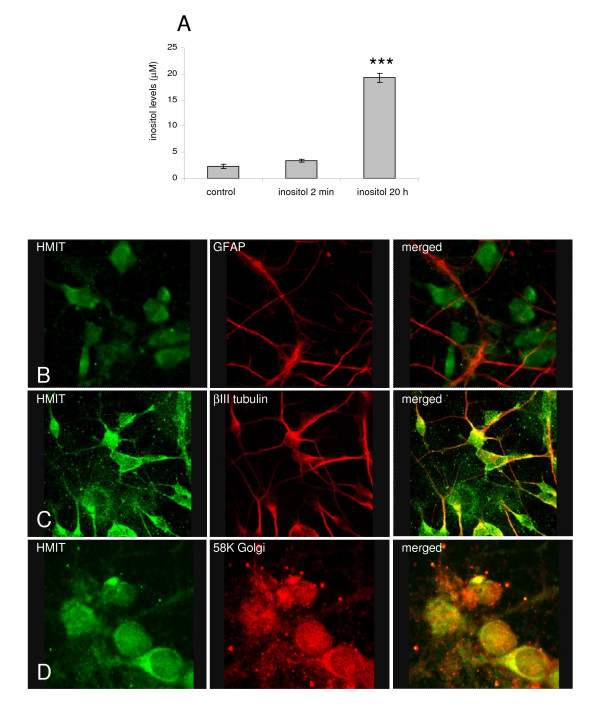
Figure 1.
Intracellular myo-inositol analysis and investigation of HMIT expression in rat dissociated neurones. A Intracellular myo-inositol levels in rat cortical neurones incubated with myo-inositol showing increased intracellular inositol levels with time (n = 3 wells from one cell preparation). Primary cortical neurones were stained with anti-HMIT:21 (1:100) in combination with: B an anti-GFAP antibody (Abcam, 1:1000); C an anti-βIII tubulin antibody (Abcam, 1:1000); or D an anti-58K Golgi antibody (Abcam, 1:1000). Secondary antibodies Alexa Fluor anti-rabbit 488 and anti-mouse 633 were used. Neurones were imaged using a confocal microscope with a 63× water immersion objective.

