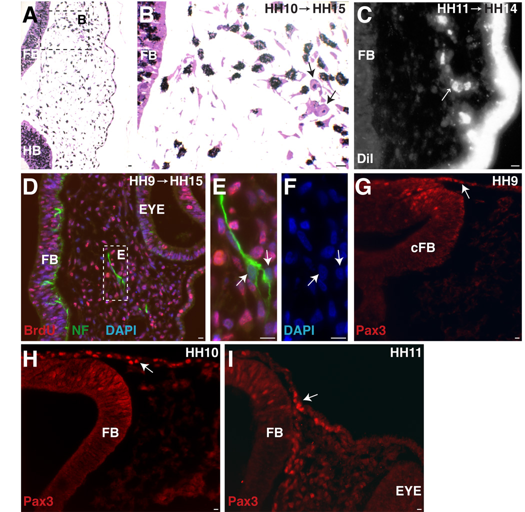
Figure 2.
Trigeminal placode extends into caudal forebrain level. A. At the forebrain/eye level, an HH10 (9ss) embryo (collected at HH15 (25ss)) shows two post-mitotic cells (arrows) that migrated to position of future opV nerve. B. Higher power of A. C. DiI label at HH11 (12ss) (collected at HH14 (21ss)) shows delaminate placode cells at forebrain level (small arrow). D. Embryo treated at HH9 (8ss) (collected at HH15 (26ss)) (same level as A,B) has two NFM+/BrdU- cells. E. Higher power of D. F. DAPI of cells in E. G–I. Pax3, a marker for specified trigeminal placode cells, is expressed at forebrain levels at HH9, 10, 11 (8ss, 11ss, 14ss). A–F. Large arrows indicate post-mitotic cells. G–I. Large arrow indicates Pax3+ cell in ectoderm. FB=forebrain, cFB=caudal forebrain, HB=hindbrain. Scale bars are approximately 10 µm. Magenta/green version (without DAPI triple label) can be found in Supplemental Figure1.

