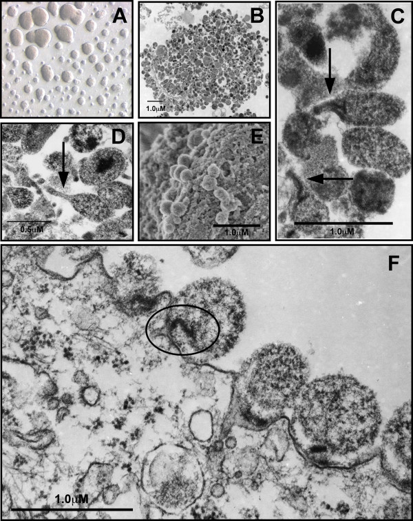
Figure 1.
Cultivation of M. genitalium and ultrastructural analysis of attachment to vaginal epithelial cells. M. genitalium G37 or M2300 were grown to log-phase in Friis FB medium. (A) Light micrograph of attached G37 microcolonies grown in culture flasks containing Friis FB medium taken using Variable Relief Contrast (VAREL). (B) TEM micrograph of a single G37 microcolony after 3d growth in Friis FB medium showing highly variable size and morphology. (C) Within M. genitalium G37 microcolonies, an elongated tip-like structure (arrow) was observed. (D) TEM micrograph M. genitalium strain M2300 showing similar variable morphology compared to G37 and formation of an electron-dense tip structure. Log-phase M. genitalium were harvested from Friis medium and then inoculated onto vaginal EC monolayers for ultrastructural analysis of attachment. (E) SEM micrograph of M. genitalium G37 attached to vaginal ECs (2 h PI). (F) TEM micrograph of M. genitalium G37 attached to vaginal ECs collected 3 h PI. An electron dense core structure presumably involved in attachment and invasion of vaginal ECs is highlighted by the oval. Similar electron dense cores were observed in some tip structures and can be seen in panel C.

