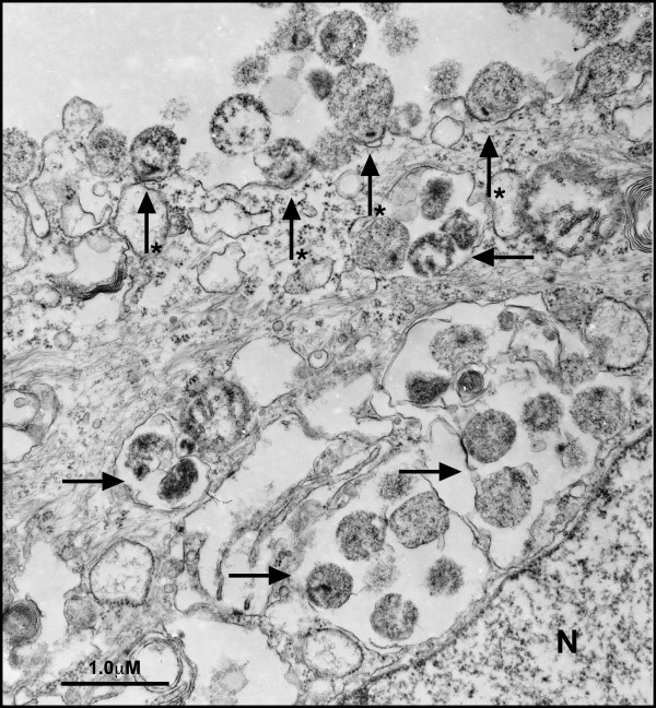
Figure 2.
Attachment and invasion of vaginal epithelial cells by M. genitalium. M. genitalium G37 or M2300 were harvested from log-phase cultures in Friis FB medium and then inoculated onto vaginal ECs. After 3 h of infection, cells were fixed and processed for TEM imaging. Many M. genitalium organisms were attached to the host cell surface associated with a polarized electron-dense core structure (starred arrow). In addition, M. genitalium organisms were localized to intracellular vacuoles (arrows) distributed throughout the cellular cytosol. Approximately 60% of observed vaginal ECs showed intracellular vacuoles directly adjacent to the nucleus (denoted as N). Similar findings were observed in cervical ECs and for the Danish M2300 strain.

