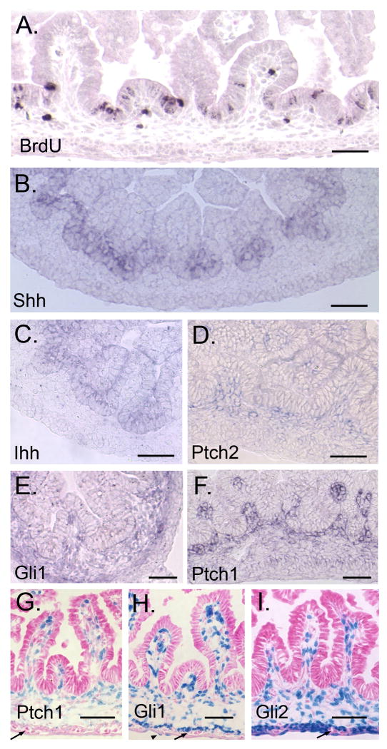
Figure 5. Hh signal transduction during villus emergence in E16.5 small intestine.
(A) BrdU staining. Epithelial proliferation is restricted to the inter-villus area. (B-F) In situ hybridization. Shh (B), but not Ihh (C) expression correlates with the restriction in epithelial proliferation. Ptch2 (D), Gli1 (E) and Ptch1 (F) are mesenchymally expressed. (G-I) X-gal staining of E16.5 PtchLacZ/+, GliLacZ/+ and Gli2LacZ/+ small intestine. All reporters are expressed in villus cores. Gli1 and Gli2 expression is also seen in ME and some serosal cells (H arrowhead). Enteric neurons are negative (arrows). Bars = 100μm (A-F) or 50μm (G-I).

