
Figure 4.
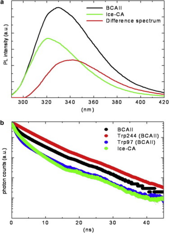
(a) BCAII and Ice-CA photoluminescence spectra obtained by exciting protein samples (2 μM in phosphate buffer, pH 7.2, at 18°C) at 280 nm wavelength, plotted together with their difference spectrum. (b) Time-resolved fluorescence emission registered at 18°C for BCAII, its Trps residues (Trp-244 and Trp-97), and Ice-CA, setting the emission wavelengths at 330, 380, 310, and 321 nm, respectively. 380- and 310-nm emission wavelengths were selected to account for the individual BCAII Trps residues (see text). λexc was 280 nm or 295 nm with no appreciable differences in the observed fluorescence decays. Ordinate scale is logarithmic.