
Figure 6.
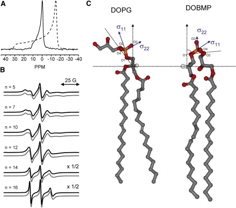
(A) 31P NMR spectrum of DOBMP dispersion (solid line) compared to POPC dispersion (dashed line), pH 7.4, T = 27 ± 1°C. Spectra are referenced to the isotropic chemical shift value of POPC. (B) Double-area normalized X-band EPR spectra for hydrated dispersions of BMP (black line) and POPC (gray line) containing 1 mol % n-doxyl spin-labeled lipid. Note that the y-axes for n = 14 and 16 are scaled by 2×. (C) Ball-and-stick model representations of DOPG and DOBMP showing the relative orientations of their phosphate head groups with respect to the bilayer normal (dotted lines). The orientation of the chemical shift tensor is taken from refs. 37, 41, and 42. Models were generated using WebLab Viewer Pro 5.0 with coordinates of individual lipid molecules with bonds rotated manually to best depict the orientation of the BMP head group proposed from molecular dynamic simulations (17) and orientations of the typical phospholipid head groups determined from NMR and x-ray models (41,42).