
Figure 1.
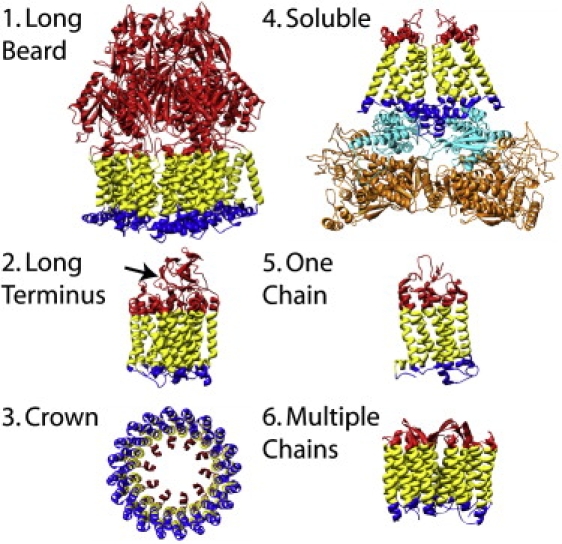
Representative structures of the groups of α-helical TM proteins defined in this article (see Materials and Methods). (1) Multidrug efflux transporter ABC (1iwg). (2) Bacterial cytochrome c oxidase (1ehk); the arrow points to the ∼120-residue-long C-terminus of chain B. (3) V-type sodium ATPase (2bl2). (4) Fumarate reductase (1l0v); chains B and N (cyan) and chains A and M (orange) are the “soluble” part of the protein complex. (5) Rhodopsin (1gzm). (6) Bacteriorhodopsin (1c3w). The membrane protein is colored as follows: yellow, TM; red, OUT; and blue, IN.