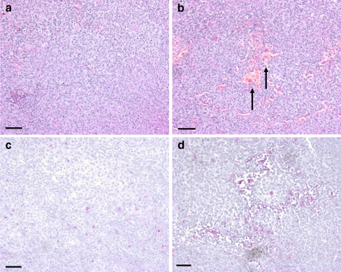
Fig. 2.
Haematoxylin and eosin staining (a and b) and immunohistochemical staining P2Y1 receptors (c and d) of excised tumour nodules. a Solid tumour from an untreated mouse. b Tumour from a mouse treated with ATP showing patchy necrosis, illustrated by red staining areas with no nuclear counterstain (arrows). c Scant expression of P2Y1 receptors (pink) in a specimen of untreated melanoma with a haematoxylin nuclear counterstain (purple). b Increased P2Y1 receptor staining in the ATP-treated group concentrated around the areas of necrosis. Haematoxylin nuclear counterstain (purple). All calibration bars = 250 µm

