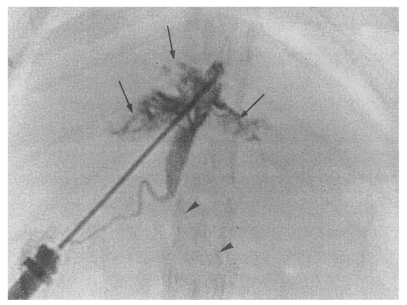
Fig. 9.
Cholangiogram of a rat in the primary infection control group 4 weeks after infection, showing moderate dilatation of the bile duct confluence and of the proximal extrahepatic bile duct. A normally appearing distal extrahepatic bile duct is also shown. Note the multiple irregular filling representing worms or desquamated material (arrows). Pancreatic duct (arrowheads).

