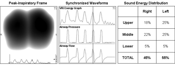
Figure 2.
An example of acoustic data as displayed for a recording obtained from a 77-year-old male with myasthenia gravis. A representative peak-inspiratory image (left panel); synchronized sound energy graph and ventilator airway pressure and flow waveforms (middle panel); sound energy distribution in the six lung regions as automatically provided by the software in percentage of weighted pixel count (right panel). VRI = vibration response imaging.

