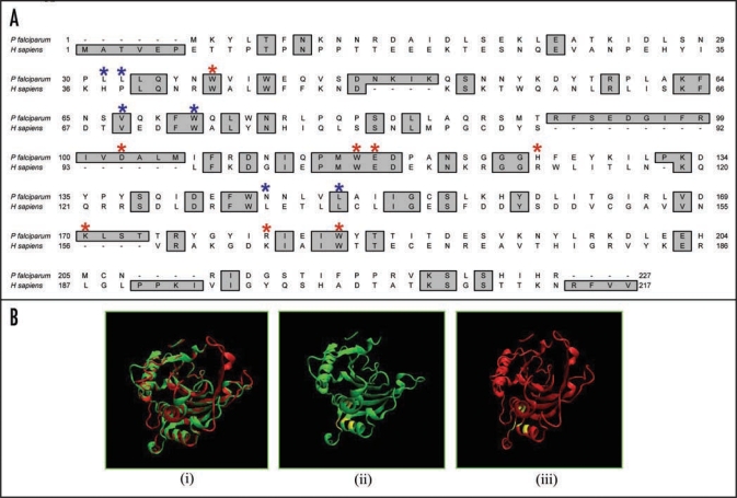
Figure 2.
(A) Amino acid alignment of P. falciparum eIF4E (PlasmoDB No. PFC0635c, GenBank accession number EF043517) and human eIF4E (GenBank accession number P06730). The amino acids responsible for 7-methyl GDP-binding and eIF4G binding are marked by red and blue asterisk respectively. (B) A three-dimensional model for PfeIF4E was created as described in text, which was based on the crystal structure of human eIF4E.16 The structures have been displayed using molecular visualization program for displaying, animating and analyzing large biomolecule systems using 3-dimensional graphics and built-in scripting (VMD software www.ks.uiuc.edu). (i) Super imposed image of the structure of human (green) and P. falciparum (red) eIF4E The amino acids responsible for the binding of eIF4G in (ii) human and (iii) P. falciparum eIF4E have been marked and are shown in yellow in the models.

