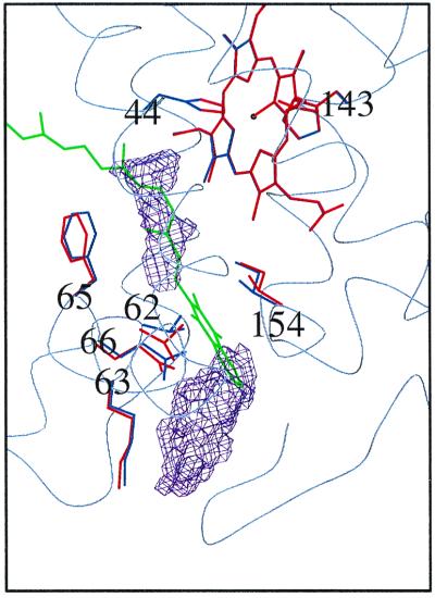
Figure 1.
A working hypothesis. Two distal cavities (purple) in subunit C of the original structure of W. succinogenes QFR (PDB entry 1QLA) as detected with the program voidoo (33) and a working model of menaquinol binding (green) are shown. To accommodate the quinol head group in its current tentative position between the cavities, amino acid side-chain movements from their original positions (blue) to positions drawn in red are required as derived from energy minimization simulations with cns. The heme group shown is the distal heme bD. In this orientation, the periplasm is at the bottom and the rest of the QFR complex extends beyond the top and the right of the figure. Figs. 1, 2, and 4 were prepared with a version of molscript (34) modified for color ramping (35) and map drawing (36) capabilities.

