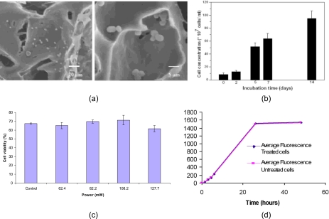
Figure 9.
(a) SEM images of yeast cells seeded into a poly(caprolactone) scaffold using SAW. The morphology of the cells do not appear to be compromised by the SAW radiation. (b) Proliferation rate of the yeast cells after irradiation with the SAW. Cells are observed to continue proliferating during the subsequent 14 days, which further confirms the viability of the yeast cells (Ref. 16). (c) Viability of primary murine osteoblast cells treated under the SAW irradiation at different rf powers. (d) Average cell proliferation of SAW-treated and untreated cells as function of the fluorescence intensity from the Alamar Blue uptake; the power of the SAW applied is 108.2 mW. In both (c) and (d), the cells were treated by the SAW at 20 MHz for 10 s, and the cell density and suspension volume is 5000 cells∕μL and 10 μL, respectively.

