
Fig. 8.
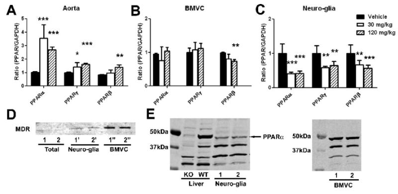
Effects of gemfibrozil on mRNA levels of PPARs in aorta (A), brain microvessels (BMVC) fraction (B), and neuro-glia fraction (C) in non-stroke mice. Mice on normal diet were treated with gemfibrozil for 7 days. mRNA levels were determined by quantitative PCR normalized by GAPDH mRNA levels in each sample. For each condition, the mean value in vehicle group was considered as 1 and relative ratio was calculated and expressed for gemfibrozil groups. *, p < 0.05; **, p < 0.01; ***, p < 0.001 by ANOVA followed by Scheffe (n = 6 or 7 in each group). D, Western blot analysis showing MDR immunoreactivity in total brain fraction, brain microvessels (BMVC), and residual fraction (Neuro-glia) containing neurons and glia. Two animals were used. E, Western blot analysis showing PPARα immunoreactivity in mouse brain. Liver tissues from PPARα knockout (KO) and wild type (WT) mouse were used to characterize the antibodies. The ∼50kDa band was weak in KO liver, suggesting that the band reflects PPARα. This band was detected in both neuro-glia fraction and brain microvessel (BMVC) fraction.