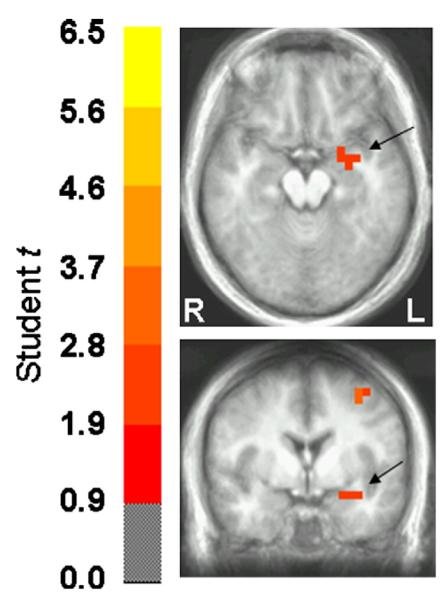
Fig. 1.

Paired t statistic volume of the threat-to-safe contrast in axial and coronal views and thresholded at p<0.05. Images are in radiological orientation (left=right, and vice versa). The center of the medial activation depicted in both views corresponds to the left amygdala according to the Talairach daemon (Lancaster et al., 2000). There was a greater left amygdala response to stimulus deviance under threat of shock relative to safe conditions between 50 and 150 ms post-stimulus onset.
