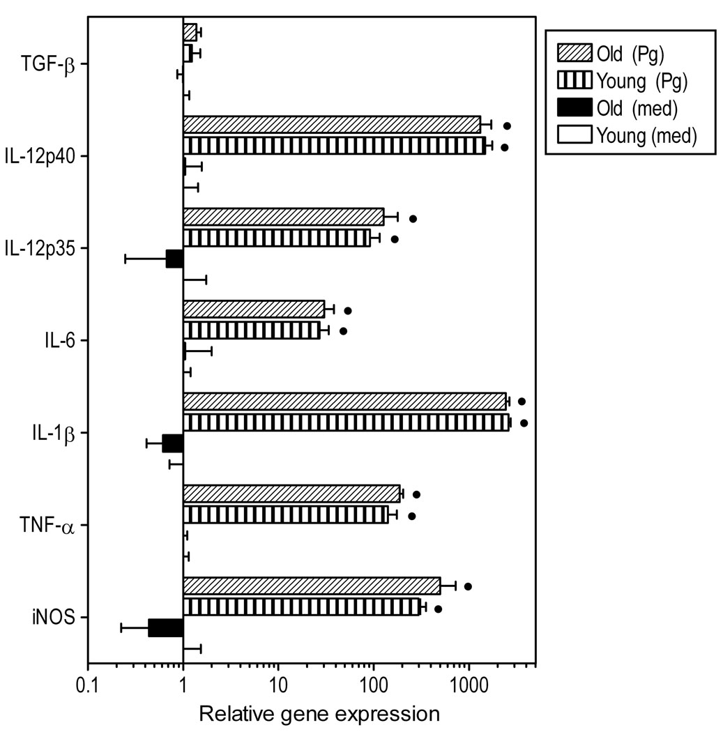
Fig. 4. Induction of innate immune response gene expression in P. gingivalis-stimulated macrophages from young or old mice.
Freshly explanted peritoneal macrophages from young or old mice were stimulated with P. gingivalis (Pg; MOI = 10:1) or medium (med) control for 4h. Quantitative real-time PCR (qPCR) was used to determine mRNA expression levels for the indicated molecules (normalized against GAPDH mRNA levels). Results are shown as fold induction relative to unstimulated “young” macrophages. Each data point represents the mean (with SD) of 5 to 10 separate expression values corresponding to qPCR analysis of total macrophage RNA from individual mice. Asterisks indicate statistically significant (p < 0.05) differences between “old” and “young” macrophages, within the same activation status. Black circles show statistically significant (p < 0.05) differences compared to unstimulated macrophages, within the same age group.

